Rewriting Life
Capturing Protein Interactions
Freezing and slicing gives a snapshot of life inside cells.
Scientists can now see how proteins organize and interact, thanks to the technology that created this image of skin tissue. The technique reveals the structure of proteins and the relationships among them in unprecedented detail, providing information that’s vital for understanding disease and cell functions. “When you see the proteins, you immediately also see … how they interact in an undisturbed environment,” says Achilleas Frangakis, the biologist who led the research at the European Molecular Biology Laboratory in Heidelberg, Germany. “At this resolution, the cell is essentially an uncharted territory.”
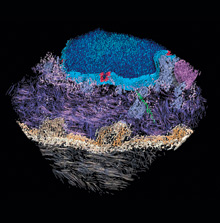
The research group froze cells to -193 ºC by plunging them into liquid nitrogen, sliced them into 50-nanometer-thick sections, and illuminated the slices with a beam of electrons. Software refined the resulting electron tomography images into virtual slices that were even thinner–as little as half a nanometer thick. Combinations of such slices enable 3-D viewing, too.
The imaging technique, called cryo-electron tomography, had previously been used on smaller, simpler cells, such as bacteria. But coupled with the slicing technique, cryosectioning, it can work on almost any cell and is “truly a first,” says Grant Jensen, a biologist at Caltech who specializes in cryo-electron tomography.