Rewriting Life
Growing Eyeballs
Embryonic stem cells growing in a dish can spontaneously form complex structures resembling the retina—a discovery that could one day help restore sight to the blind.
A clump of mouse embryonic stem cells can self-organize into three-dimensional structures reminiscent of the retina in the early stages of embryonic development, according to a new study published Wednesday in Nature. Researchers believe this process could one day serve as a source of cells to transplant into diseased and damaged retinas—a potential way to restore sight to the blind.
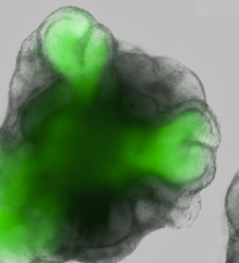
Researchers at the RIKEN Center for Developmental Biology in Kobe, Japan, began with clusters of about 3,000 mouse embryonic stem cells floating in a mix of chemicals designed to spur differentiation into retinal cells. After a week, several balloon-like sacs of cells began to protrude from the surface of each cluster. Over the next few days, those sacs pouched inward on themselves to form structures resembling the optic cup—the complex dual-layered structure that emerges early in development and eventually becomes the retina.
This was surprising because the cells were not coaxed in any way to form such a structure, says Yoshiki Sasai, director of the Organogenesis and Neurogenesis Group at RIKEN and lead author of the study. From a homogeneous mass of embryonic stem cells, a sophisticated three-dimensional tissue spontaneously emerged. In other words, says Sasai, “the shape of an organ is actually internally programmed.” This concept could have profound implications for the field of stem cell research; Sasai suspects that self-organization is possible for other tissue types as well. If that’s true, scientists could potentially grow many kinds of organs in the laboratory.
“I think it really represents a significant step forward,” says Jane Sowden, a developmental biologist at University College London. Sowden was not involved in the study. “Previous studies have shown that it’s possible to differentiate [embryonic stem] cells into distinct types of retinal cells, but what we’re seeing in this study is the potential of these cells to organize themselves into tissue of the retina.”
The research may also have a more immediate impact on treatments for diseases in which the photoreceptor cells of the eye are damaged or destroyed. Sowden’s group has found that transplanting photoreceptor precursors—a type of cell that appears early in development—could restore sight in these cases. But it has been difficult to obtain large numbers of these cells. Because the structures in the new study developed according to a predictable pattern and timeline, Sowden says, they could provide an ideal source of photoreceptor precursors for transplantation.
Sasai also believes the entire inner sheath of the optic cup-like structure—which contains photoreceptor cells arranged in the particular intricate layout that is necessary for sight—could be transplanted. His group has already had some success with this approach in mice, and they hope to have a human version of the system within two years.
“The ability to replicate organogenesis in the lab could revolutionize stem cell medicine,” says Robert Lanza, chief scientific officer at Advanced Cell Technology. Lanza was not involved in the study. “By understanding the necessary signals and forces, we can hopefully develop similar culture systems to encourage the self-directed organization of other complex tissue and organ systems needed for transplantation.”