
A Renaissance Woman for the Nano Age
Sangeeta Bhatia, SM ’93, PhD ’97—an engineer, doctor, entrepreneur, and mother—invented a device that tests whether potential drugs are toxic. Now she’s working on fighting malaria and curing cancer.

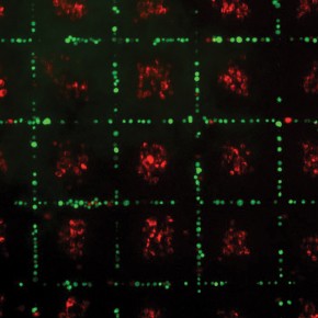
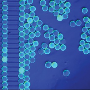
As a graduate student in the Harvard-MIT Division of Health Sciences and Technology, Sangeeta Bhatia figured out a way to keep liver cells alive outside the body, sustaining their function for weeks at a time. Working with microfabricated surfaces similar to those used for computer chips, she arranged the finicky cells in patterns of stripes and dots. Adding nutrients and other cell types, she arrived at an architecture that made the collections of cells function like tiny livers, metabolizing drugs and producing proteins. Her breakthrough model and its successors have allowed Bhatia, now a bioengineering professor at MIT, to investigate drug toxicity and metabolism as well as a host of diseases that affect the liver.
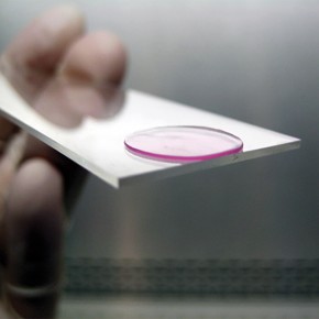
“She didn’t just stop at saying, ‘I can grow some cells in a dish and they function better,’” says Tejal Desai, a professor and chair of the department of bioengineering and therapeutic sciences at the University of California, San Francisco. Instead, she “pushed our understanding all the way from the basic science” into clinical applications. Pharmaceutical companies around the world, she notes, use a device based on Bhatia’s microlivers to test the toxicity of potential drugs and study how they’re metabolized.
Bhatia’s laboratory also works on projects aimed at using targeted nanoparticles to diagnose and treat cancer. Even in a field known for interdisciplinary work, she has an unusual knack for integrating ideas from different areas, including sensor technology, chemical biology, nanotech, and engineering, says Desai.
Bhatia was born in Boston, to immigrant parents. Her mother, she says, was one of the first female MBAs in India. Her father was an engineer. Traditional in their outlook, Bhatia’s parents thought the three acceptable jobs for their daughter were engineer, doctor, and entrepreneur. They now laugh about the fact that, as she puts it, “I’ve become all three.” (In fact, Bhatia’s formal titles at MIT alone stretch the limits of a business card: she’s director of the Laboratory for Multiscale Regenerative Technologies, the John J. and Dorothy Wilson Professor of Health Sciences and Technology & Electrical Engineering and Computer Science, and a member of both the Institute for Medical Engineering and Science and the Koch Institute for Integrative Cancer Research. And that’s to say nothing of her affiliations with the Broad Institute, the Howard Hughes Medical Institute, the Ludwig Center for Molecular Oncology, the Harvard Stem Cell Institute, and Brigham and Women’s Hospital.)
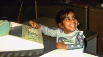
From an early age, Bhatia was a natural tinkerer. She once took apart the family’s broken answering machine, decided that a few parts seemed out of place, and put it back together again, restoring its function. In high school, she gravitated toward biology, and her father encouraged her to think about biomedical engineering, then a nascent field. During college, at Brown University, Bhatia explored work on nerve regeneration, excited to focus on projects aimed at promoting health.
In 1991, after a brief stint at a pharmaceutical company, Bhatia enrolled in a mechanical engineering program at MIT. She came “running back to grad school,” she recalls, because her job in pharma felt “too far from the human interface.” Eventually, she would join the lab of Mehmet Toner, SM ’85, PhD ’89, a young biomedical engineer in Mass. General Hospital’s department of surgery whose enthusiasm “just bowled me over,” she says. Toner’s goal was to create a device analogous to a dialysis machine for patients with acute liver failure: liver cells in the machine would process patients’ blood before returning it to the body. Bhatia’s challenge was to find a way to sustain the cells within a mechanical cartridge.
This proved difficult. Working with animal cells, Bhatia tried to re-create the architecture of the liver by arranging them on a pattern of water-attracting and water-repelling surfaces. After two years of failed efforts, though, she realized she had hit a dead end. “It just wasn’t going to happen,” she says. At the suggestion of a classmate, she eventually teamed up with the MIT microfabrication facility, which had, until then, focused almost exclusively on making computer chips. Bhatia’s project, involving a chip for a cellular study, was one of the first of its kind at MIT.
By this time, Bhatia was enrolled in the Harvard-MIT Division of Health Sciences and Technology. (She transferred in, having been rejected from the program the first time around, she recalls with a wry smile.) As part of the HST program, she took medical school classes and found herself “falling in love with the human body.” She decided to complete an MD, but she couldn’t resist throwing her hat in the ring for research opportunities too; and during her third year of medical school, she got an offer from the University of California, San Diego, to join the faculty. Remarkably, Bhatia completed her fourth year of medical school in San Diego while taking on the responsibilities of a junior professor. “I worked in the hospital by day, doing rotations, and set up the lab at night,” she says. And though the tenure clock was ticking for a year before she could devote herself fully to research, she was appointed an associate professor at UCSD in 2002.
A few years later, however, she was ready to return to the East Coast. She and her husband, Jagesh Shah, SM ’95, PhD ’99, whom she’d met on her first day at HST and who had moved with her to California, had had a daughter in 2003. Living closer to extended family in Boston and Toronto became a priority: it was “a big part of the life I always wanted,” she says. So she and Shah conducted a joint search, applying for positions together or at neighboring institutions. Upon receiving twin offers from MIT and Harvard, they moved to the Boston area in 2005.
Meanwhile, Bhatia’s work on microlivers surged forward. She began using human rather than animal cells, and in a set of pivotal experiments, she showed that her system could flag pharmaceutical compounds that might pose a danger to people. For instance, a drug called fialuridine, which had been investigated in the 1990s as a therapy for hepatitis B, had gone through animal testing without raising safety concerns. But in clinical trials, it caused liver failure and death in five subjects. Bhatia showed that her system would have identified fialuridine as unsafe. In 2008 she cofounded a company called Hepregen to manufacture the liver-chip device and distribute it to pharmaceutical companies. Today, over 40 companies around the world use Bhatia’s method to test drugs’ safety in liver cells before doing clinical trials. “We can catch drugs that might not otherwise be caught this early,” she says. Bhatia and colleagues have also used her models to study diseases affecting the liver, such as hepatitis C, hepatitis B, and malaria.
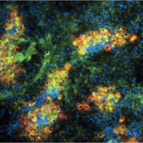
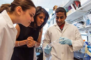
On a sunny October afternoon, in one of her lab’s six sprawling rooms, Bhatia points to small bins where she and her team store malarial mosquitoes, infected with blood that Nil Gural, an HST graduate student in the lab, collected from patients in Thailand. To do their research, Gural and others cut the mosquitoes’ heads off and remove their salivary glands, which are filled with parasites. Then they add these parasites to a culture of liver cells. Their goal is to monitor the progress of infection and, specifically, to study what happens when the parasite becomes quiescent, which typically happens after about three weeks. This is how malaria hides in the liver, Bhatia explains, and it is probably “the biggest barrier to eradicating the disease” once it’s established in the body. The dormant form of the parasite has also been challenging for researchers to study, because liver cells are so difficult to keep alive outside the body. Since 2009, however, Bhatia’s group has used her model system, in which liver cells can survive for up to a month, to study the process by which the parasite becomes inactive. Ultimately, Bhatia hopes, this work will shed light on how exactly the dormant pathogen becomes activated again in patients. She also hopes it will help researchers test drugs aimed at vanquishing the parasite entirely.
Elsewhere in the lab, a range of equipment attests to the breadth of Bhatia’s research program. There’s a 3-D printer for prototyping, an anesthesia bay for small animals, and a magnetic robot that is used to levitate magnetic nanoparticles. “This is Maggie,” she says, affectionately patting the long robotic arm. In addition to working on the liver, Bhatia’s group, which is located in the Koch Institute for Integrative Cancer Research, investigates ways to direct nanomaterials to tumors in order to image, diagnose, and ultimately treat cancer. This possibility has been an interest of Bhatia’s since early in her career. In 2000, she began collaborating with cancer researcher Erkki Ruoslahti of the University of California, Santa Barbara, who had developed peptides that bind to the markers that tumor blood vessels produce. These peptides can be attached to the surfaces of various materials to home in on tumors. Bhatia and Ruoslahti also worked closely with materials scientist Michael Sailor at UCSD. In one project, the group focused on directing contrast agents to tumors in order to enhance imaging technologies. With their technology, materials such as nanoparticles of iron oxide, which is used in magnetic resonance imaging, would go directly to the cancer, allowing a surgeon to see more clearly whether, say, a lymph node had been infiltrated by disease.
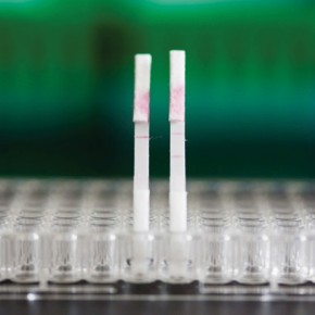
Over the years, Bhatia, Sailor, and Ruoslahti have continued a fruitful collaboration. “We’ve been a three-way team,” says Sailor, adding that he and Bhatia “hit it off immediately” and brainstormed a dozen possible projects the first time they met, in the late 1990s. In more recent work, the team has focused on directing nanomaterials to tumors that are metastasizing. They introduced peptides that would be cut by the tumors’ enzymes and would also produce fluorescence. And while the original intent was to highlight the tumor for MRI scans, they soon noticed that “the bladder of every animal with cancer was lighting up with fluorescence,” says Bhatia. As it turned out, the way the materials interacted with the enzymes was creating a small fluorescent biomarker that was entering the kidneys and being released in urine. “We had a big ‘Aha’ moment where we realized we don’t need the MRI machine at all,” says Bhatia. Instead, they could just test urine for telltale biomarkers of cancer. In additional work, the team played with several ways these markers could be detected in urine. For instance, they created a paper-based test, similar to the kind used for pregnancy, that could flag the presence of tumors. Recently, Bhatia cofounded a company called Glympse Bio, aimed at commercializing the technology. Ultimately, the goal would be to develop tools to detect early-stage cancer. Although she describes such tools as a “dream” and cautions that they’re a long way off, she hopes that they one day might include organ-specific tests, analogous to mammography for breast cancer or colonoscopy for colon cancer, that would “detect cancer before current tests would be able to.”
Most of Bhatia’s projects on medical uses of nanomaterials have involved injecting the materials into animals. But last year her lab and collaborators at UCSD also found a novel way to deliver these cancer-detecting materials: in bacteria that could be consumed in yogurt. When these modified bacteria entered the animals’ bodies, this time by way of the GI tract, they once again homed in on tumors and created biomarkers that could be detected in urine. This approach will probably work best for tumors of the GI tract, including the colon, since it’s right there, says Bhatia. It might work well for detecting liver tumors, too, since nutrients and metabolites reach that organ relatively directly. In another project, Bhatia is using modified bacteria to treat cancer rather than simply diagnose it. There, the goal is for bacteria to zero in on tumors and deliver molecules that will kill them entirely. “We call this our programmable probiotic project,” she says.
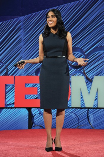
Bhatia, 47, has accrued an impressive list of honors and awards, including the Lemelson-MIT Prize, the Packard Fellowship, and the Heinz Award. She is also a member of the American Academy of Arts and Sciences and the National Academy of Engineering. Yet even as her career soars, she is mindful of the challenges faced by female scientists and devotes considerable time to supporting others. “When I started at Brown [in the mid-1980s], my best friend and I looked around in our freshman engineering class and saw 50 percent men and 50 percent women and thought, ‘What is all the fuss about?’” she says. They figured it was “just a matter of time” before women would be well represented. Yet by the time she graduated, only 16 percent of the engineers in her class were women.
This attrition is what social scientists call “the leaky pipeline.” And for Bhatia, getting girls interested in science—and being an active role model to encourage them to stick with it—has long been a passion. She made time to organize (with Shah) the science fair at their daughters’ elementary school for several years. As a grad student, she helped start a program called Keys to Empowering Youth, which brings middle school girls to MIT each year to participate in lab work. That experience can boost their confidence early on, perhaps increasing the chances they will choose to study science and lending them the resilience to persevere in scientific careers. She also tries to make herself publicly visible, especially to young women. “My natural tendency is to want to be in the lab and do my work, but I feel you can’t be a role model if people can’t see you,” she says. In press appearances, she comes across as grounded and relatable as well as scientifically serious. A 2009 Nova profile, for instance, showed her two daughters doing science experiments with her in the kitchen, going on a shopping trip for toys (but not Barbies), and jumping into bed with her and her husband in the morning—a rare glimpse into the personal life of a senior scientist. She talks about weekly date nights with Shah. And college friend Theresia Gouw (now a venture capitalist) confirms that Bhatia keeps in close touch with her friends; an annual girls’ weekend usually involves shopping and spas. “I try to be pretty open about having a life, having children and a rewarding career, so hopefully women will want to choose this,” Bhatia says.
In her lab, as well, she fosters a supportive intellectual camaraderie. “She’s good at creating an environment where people actually want to help each other and collaborate,” says graduate student Gural. Bhatia also serves as advisor to the MIT Society of Women Engineers and advocates for female scientists both at MIT and elsewhere. “For me it’s an obligation to raise awareness,” she says, citing unconscious bias against female researchers in hiring, promotion, and speaking opportunities as an issue some scientists may still not be aware of. Nationally, only around a quarter of engineering doctorates are earned by women, roughly 15 percent of tenured or tenure-track engineering faculty are female, and only a tiny fraction of tech startups have female founders. “People still say it’s a matter of time, and it is getting better,” she says. “But I’m pretty impatient about the slope of the line.”

Advertisement