Intelligent Machines
Remaking X-Rays
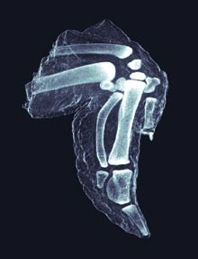
Silicon gratings heighten contrast.
The basic physics behind x‑ray imaging haven’t changed in more than 100 years. While most hospitals and airports have gotten rid of film and gone digital, their systems still record how much x-ray radiation passes through your arm or your suitcase and how much is absorbed. But the bones in your arm don’t just absorb x-rays; they also “scatter” them, or deflect them from their paths. Those scattered x‑rays could yield valuable information, but they tend to get drowned out by the stronger signal from unscattered rays. Now Franz Pfeiffer, assistant professor of physics at the École Polytechnique Fédérale de Lausanne, and Christian David at the Paul Scherrer Institut in Villigen, Switzerland, have created grooved silicon gratings that filter out much of the unscattered radiation, so the signal from the scattered rays is clearer. The resulting images could provide enough extra detail to reveal smaller tumors or distinguish a block of explosives from a chunk of cheese.
Pfeiffer demonstrated the gratings in the lab, adding them to conventional x-ray tubes; now he’s trying to incorporate them into hospital CT scanners, which use x-rays. The researchers are collaborating with others to determine whether oncologists might use the new technique to produce higher-contrast mammograms with a lower false-positive rate.
