Rewriting Life
Superthin 3-D Endoscope
An instrument to detect tiny tumors
In an endoscope meant to penetrate the brain, look at a fetus, or thread through tiny ducts, smaller is better. But the endoscopes that produce the clearest 3-D images use cameras several millimeters wide–too big to go many places in the body. Now researchers at Massachusetts General Hospital in Boston have demonstrated an endoscope that’s just 350 micrometers wide and sends back 3‑D images that are as clear as those produced by larger endoscopes.
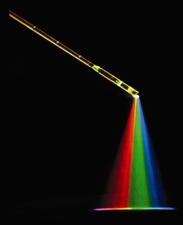
The key to the device is how it uses light, says Guillermo Tearney, professor of pathology at Harvard Medical School and the project leader. In the endoscope, white light moves down a glass fiber and is broken into a rainbow of colors by an optical device called a diffraction grating. Each color hits a different part of the tissue being imaged, reflects back, and travels through the fiber to a spectrometer outside the patient’s body. Each color provides a separate pixel of information. A computer compares the reflections with a reference beam to create a 3-D topography.
The scope’s scans of a mouse abdomen showed 100-micrometer tumors on the abdominal wall. If doctors could see tumors that small in humans, they might catch cases of breast, pancreatic, and other types of cancer sooner, Tearney says. The device could also make it possible to perform new types of brain surgery and fetal surgery.
The new endoscope, still a prototype, must undergo safety testing before reaching humans, Tearney says, and it provides only slightly better resolution than existing scopes. But new versions in the works might feature improvements that boost resolution tenfold.