Rewriting Life
Finding the Core of the Brain
A new mapping approach identifies the hub of the human cortex.
The iconic image of the brain is a misshapen, yellowish lump. Existing technology can show which parts of the lump light up when people think, but a real understanding of how the brain works demands a better picture of the nerve fibers that ferry electrical signals between brain cells. Those fibers, however, are so small and tangled that researchers haven’t been able to see them clearly.
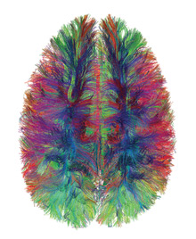
Now, an international team of scientists has combined a new variation on magnetic resonance imaging with mathematical analysis to generate the first detailed map of the network of connections in the human cortex, the part of the brain responsible for higher-order thinking.
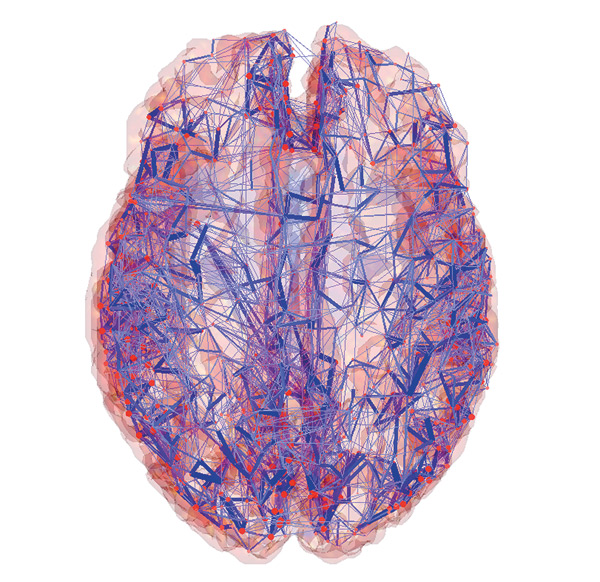
Diffusion spectrum imaging–which tracks water molecules moving along nerve fibers–gave the scientists a wiring map of the cortex, revealing points where multiple nerve fibers converged. The scientists then used a mathematical technique to repeatedly prune away the connection points with the fewest links. “If you do it gradually, you end up with a set of nodes remaining that are highly interconnected,” says Olaf Sporns, the Indiana University researcher who performed the analysis.
The most highly connected node is at the back of the head, and it lies on the shortest path between many different parts of the neural network. Not only does it have many internal connections, says Sporns, but it’s “highly central with respect to the rest of the brain.”
The researchers want to use the imaging technique to look at conditions such as schizophrenia, autism, and Alzheimer’s disease, all of which have been linked to disturbances in brain architecture. “We would like to know where the disturbances are and whether we can understand something about the clinical condition based on the connectivity,” says Sporns
The network of connections in the cortex.
Credit: Van J. Wedeen, Patric Hagmann, Olaf Sporns
