Generating cells from patients suffering from such disorders as Down syndrome, schizophrenia, and ALS lets scientist study these diseases in the lab and test new drugs.
Just four years ago, scientists from the United States and Japan developed a way to create stem cells from human skin cells. Since then, researchers have used the technology to generate such cells from people with a variety of different diseases, including diabetes, Down syndrome, and Parkinson’s disease. By differentiating these cells into the cell type affected in the disease, scientists can search for molecular missteps unique to these cells. The findings are already beginning to shed light on these diseases and are being used as a tool to test new treatments.
Here, researchers created stem cells from patients with a rare neurological disease, familial dysautonomia, and then differentiated them into the specific neurons (shown labeled with red and blue markers) affected by the disease. They found that the cells did not differentiate into neurons as readily as cells derived from healthy people, nor did they migrate as easily as normal cells. Researchers used the cells to test a handful of potential treatments for the disorder, identifying one candidate that reversed the defect in differentiation.
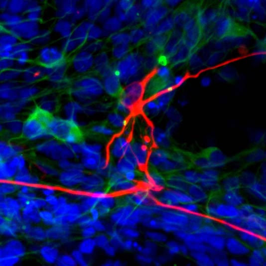
Ricardo Dolmetsch and collaborators from Stanford created neurons from a patient with Timothy syndrome, a rare inherited disorder that affects both the heart and the brain and can cause autism. Growing patient-derived neurons (above, red) in a dish, the scientists discovered that the cells were making too much of an enzyme (shown in green) that is critical for producing dopamine and norepinephrine, two important chemical messengers in the brain. Treating the cells with a specific drug blocked some of the excess.
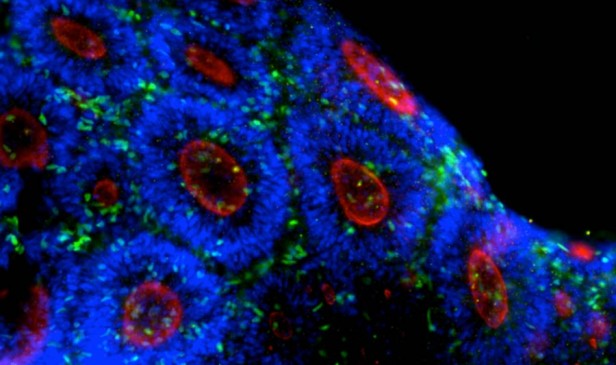
Here, neural precursor cells grown in a dish form shapes similar to the neural tube, a structure formed early in brain development. Cells derived from patients with Timothy syndrome have less of the molecule n-cadherin (shown in red), which helps cells stick together. This reinforces the view that autism is linked to defects in brain connectivity. The findings were published Sunday in Nature Medicine.
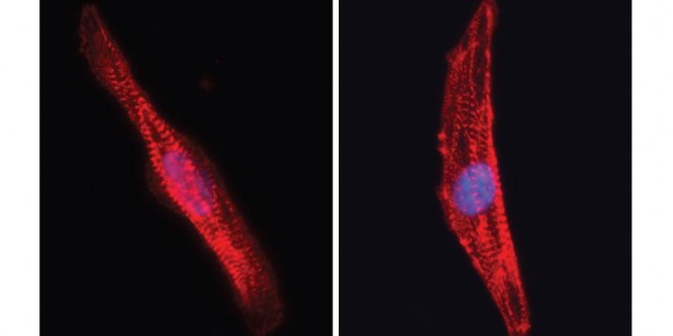
Dolmetsch and collaborators had previously created heart muscle cells (red) from people with Timothy syndrome, revealing that cells created from affected people (right) had a greater concentration of calcium (blue) than did heart muscle derived from healthy people (left).
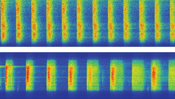
While heart cells from healthy people (top) beat at a regular 60 beats per minute, cells from patients with Timothy syndrome (bottom) beat more slowly and miss certain beats. Researchers have already identified one compound that normalizes heart rhythms in cells growing in a dish.
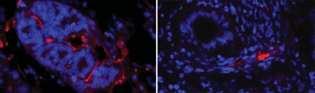
Studying stem cells derived from people with Down syndrome (right) reveals a possible reason that those with the disorder have dramatically lower rates for certain types of cancer; these cells generate fewer blood vessels (shown in red) than those derived from a chromosomally normal individual (left).
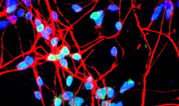
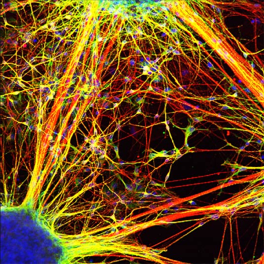
Neurons derived from schizophrenia patients form fewer connections than those from people without the disease. Cell nuclei are shown in blue, and branched fibers connecting neurons are green and red.
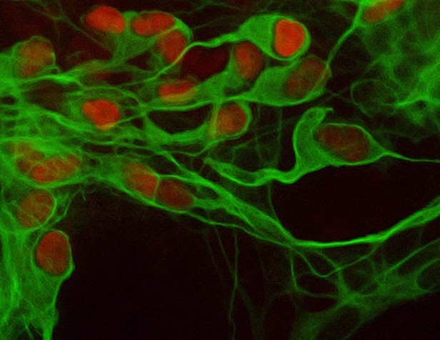
Scientists generated motor neurons (the cell nuclei are shown here in red), which are destroyed in amyotrophic lateral sclerosis (ALS), from stem cells created from a patient with the disease. The newly created cells should allow scientists to study the disease and screen new drugs. Neurons are marked in green.
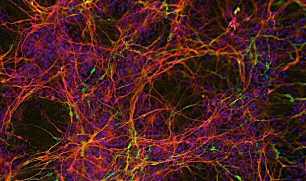
Scientists generated a type of nerve cell called an astrocyte (shown here in red) from stem cells created from a patient with ALS. Previous studies suggest that these cells play an important role in the progression of the motor neuron degeneration. The green cells are neurons, while the blue circles highlight cell nuclei.
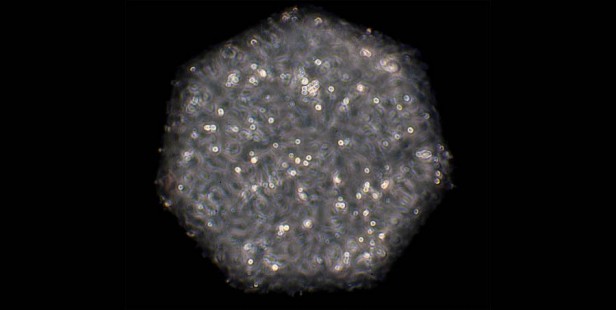
Technology Review contributor David Ewing Duncan had iPS cells created from his own tissue that were differentiated into a beating clump of heart cells (shown here). This approach might one day be used to customize drug regimens for individual patients.